Thermography is a noninvasive, safe, and affordable form of diagnostic imaging that's a helpful tool in equine health care.
Like other technologies, we're currently seeing significant advancements in cameras' resolution and user-friendliness, together with substantial drops in initial purchase costs and physical size.
As thermography gains popularity and interest, it's crucial that technicians veterinarians and horse owners know the benefits this type of imaging provides, as well as its limitations.
The first step in knowing what thermography can and can not do is learning the difference between physiologic and anatomic imaging.
Anatomic imaging, which includes MRI, CT scans, and radiographs, illustrates a particular affected area. These methods provide us with a snapshot, one moment in time, of a body part and are not sensitive to metabolic changes in nerve conduction or blood flow.
Physiologic imaging, on the other hand, is sensitive and dynamic and includes both nuclear scintigraphy (bone scan) and infrared thermography. IR imaging can detect metabolic changes associated with blood circulation, active inflammation, or nerve conduction. Veterinarians generally follow thermography with anatomic imaging to analyze the underlying disease process at a specific area of activity or concern.
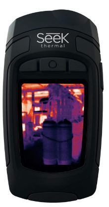
A thermographic camera identifies infrared waves on the body surface that the human eye can't see and transforms them into a picture we can see.
Suppose what happens if you injure yourself: You hit your knee; the area becomes red, inflamed, and hot; and at a cellular level as an immune response, the body releases chemicals like histamine.
Variations in blood flow might directly associate with inflammation. Therefore, at the most basic level, where there's increased circulation, there might be inflammation.
The reverse is also true: with chronic disease, nerve damage, scarring, atrophy (muscle wasting/loss), or disuse, areas might become cooler.
Symmetry is critical when interpreting images. By comparing the components of a horse left-to-right, the specific patient functions as its own control.
Additionally, consider that a horse with an injured right front flexor muscle, for example, might be eliminating pressure from the painful leg to the opposite limb or even the hind end.
With a complete horse scan, veterinarians can find these compensatory issues, start treatment, and prevent further injury.
Performance-limiting or career-ending conditions like tendon/ligament problems, kissing spines, and soreness from poorly fitted saddles, arthritis, hoof abnormalities, and cervical injuries all make patterning detectable with thermographic cameras.
Thermography has a wide range of applications such as saddle-fitting evaluation, shoeing and trimming assessment, lameness localization, serial assessment of soft-tissue healing, arthritis detection, pre- and post-chiropractic or acupuncture or massage treatment/evaluation, tendon/ligament evaluation, etc.
Researchers have shown they can use thermography to identify tendon/ligament injuries up to two weeks before evidence of clinical lameness, and subclinical hock inflammation before bony changes.
Scientists use thermography in research settings to assess physiologic responses to drugs, equipment such as equine treadmills, and diseases of the hoof. It's essential to remember that the camera reads surface temperatures and does not penetrate; therefore, veterinarians cannot assess deeper structures.
Environmental factors and patient preparation and positioning are essential for maximal results.
Exposure to radiant heat, sunlight, moisture, and wind can ruin a scan and wreck image and interpretation quality.
Also consider additional insulating factors, such as feathers, blankets, long hair coats, or boots, when making the patient ready.
When veterinarians use a standardized strategy for preparation and imaging can be a tool for care.